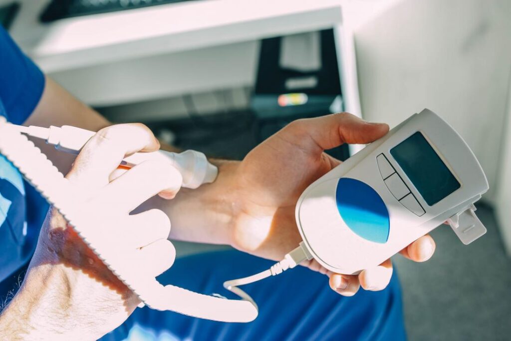
The Doppler is an examination that allows you to explore the blood flow in the arteries and veins. Its purpose is to detect the possible presence of an obstacle that would prevent proper irrigation of the organs. It is prescribed in case of suspicion of phlebitis or atheroma plaques.
What is the Doppler used for?
- The Doppler studies the flow of blood in the arteries and veins, thus giving information on its flow conditions and the proper irrigation of the organs. Coupled with ultrasound, it provides information on the shape of the vessels.
- This examination can be used on the vessels of the limbs, neck, abdomen …
- He looks for disturbances in blood flow that may be related to an obstacle or a narrowing of the vessel.
- It may be a blocked clot in a vein (this is the phlebitis), a narrowing of the caliber of an artery (these are atheroma plaques) …

What is a Doppler?
Doppler uses ultrasound, not x-rays. It works on the same bases as ultrasound, with which it is very often associated.
Its principle consists in studying the flow of blood in a vessel. A pen-shaped probe emitting ultrasound is applied to the region to be examined. The ultrasound wave propagates in the tissues and is returned in the form of an echo by the various organs it encounters. This signal is analyzed and transformed into a sound, a curve or a color reflecting the speeds of blood circulation.
The Doppler system is most often integrated into the ultrasound machine. The room where the Doppler is performed consists of:
- The probe that emits the ultrasounds and collects the signal after it has passed through the tissues. It is connected to the device by a cable.
- Video screen on which the images are viewed.
- The computer system.
- The control panel consists of multiple keys and functions.
There are three different types of Doppler:
- The continuous Doppler which translates the speed of blood flow into a sound that can be analyzed by the ear of the radiologist during the examination.
- Pulsed Doppler which translates velocity into a graph.
- The color Doppler which is associated with ultrasound to give an image of the vessel colored by blue or red depending on the direction of blood circulation.

How is a Doppler performed?
- This examination is carried out by a specialist in radiology.
- After reporting your arrival at reception, you will wait a few minutes in the waiting room.
- Before the exam, you will go to the cloakroom to undress (you will be told which clothes to take off).
- Do not forget to go to the toilet for more comfort.
- During the exam, you will lie on a bed, usually on your back.
- A gel will be spread on the skin to allow good transmission of ultrasound. The probe (like a pen) will be moved next to the vessels to be explored. The radiologist will ask you to change position during the examination and/or hold your breath.
- It takes about 10 to 20 minutes. It’s a quick review!
- The results: the radiologist will give you an initial comment. The final report will be sent to your attending physician as soon as possible. He will explain the results to you and tell you what to do.
Is the Doppler painful?
It is a completely painless examination.

How to prepare for a Doppler?
- No preparation is necessary.
- No need to fast. You can eat and drink normally.
- Consider bringing:
- The letter from your doctor and your prescriptions.
- Your social insurance cards.
- Your old shots that will allow a comparison.
What are the risks of a Doppler?
There are not any. Ultrasounds are harmless.


