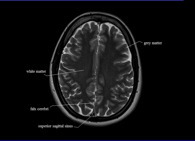
What is an MRI brain scanner and how does it work?
The MRI scanner is basically a piece of equipment that studies the magnetic properties of tissues. Each tissue has different magnetic parameters, and, more importantly, these parameters normally vary when there is an injury. This fact allows the team to make different types of images, assigning a specific gray scale to the different magnetic parameters that it measures.
The advantage of having the possibility of obtaining different contrasts is that it allows for detecting a greater number of lesions, those that are invisible in one type of contrast stand out in another, as is the case with this ischemic lesion due to blockage of an artery:
brain magnetic resonance imaging lesions
Therefore, a brain MRI scan usually consists of several images, each of which is acquired in between 2 and 8 minutes. Hence the total duration of the scan.
But how does the teamwork?
To study the magnetic properties of tissues we need a magnetic field and, for this, a magnet. In short, it is what the tube is: a huge electromagnet (electric current rotating through a circuit that generates a magnetic field) with enough force to lift a car.

For this reason, MRI is contraindicated in people with pacemakers (it could stop) and other metallic, electronic magnetic devices. When the person is introduced into the tube, each of their tissues is magnetized according to their characteristics, very little but enough to be detected and to carry out the study. The team searches for the MRI of the tissues with radio waves.
This is another advantage of magnetic resonance imaging: it does not use X-rays or ionizing radiation, only radio waves in the region of the modulated frequency (FM from 63 to 126 MHz, depending on the team) that are harmless.
Magnetic resonance makes the magnetization of the tissues vary in direction. When the magnetic resonance ceases, magnetic relaxation occurs, the way back to the starting position, each tissue at its own speed. The magnetic relaxation in turn generates a wave that is picked up by an antenna. The information from the wave is sent to a computer that reconstructs the layout of the tissues, thereby generating the image.
Brain MRI as a research tool
Since its inception, magnetic resonance imaging has aroused the interest of scientists. For Dr. Falcón “its versatility allows obtaining not only anatomical images in different contrasts, but also dynamic and functional images, extremely useful information in understanding the functioning of the brain and the processes involved in any pathology. What was previously only could verify in post-mortem studies it is now visible in vivo with scans, somewhat annoying but innocuous, of just under an hour” for more info visit labuncle.com.
The main difference between MRI used for diagnosis or as a research tool is the precision, much higher in the case of research, and often the type of information that is obtained from the tissues. Let’s review some research explorations in neuroimaging with magnetic resonance:
1) Anatomical images
The objective is to know the anatomy of the brain with maximum precision, to take reliable measurements that can be monitored over time. The difference with equivalent diagnostic images is only the resolution.
Below is an example of a diagnostic image (left), its equivalent in research (center) and super-resolution (right). The first is sufficient to rule out pathology, but is it the quantitative measurements would be inaccurate due to the blurring of the edges.
2) Anatomical images without diagnostic value
This case would be, for example, the study of white matter tracts that inform which part of the brain is connected to which other part and the strength of these connections (an example below). This information allows us to understand the network structure of the brain and its functioning when performing different tasks.
3) Functional images
Complementary to the previous one. It determines which part of the brain is activated or deactivated when performing a certain task and allows us to understand how the brain works (below, the activation when listening to emotional music, with the activation of the auditory -lateral-, emotional -central- and frontal -the superior cortex -) and what goes wrong when there is mental dysfunction. They also make it possible to determine which parts of the brain work synchronously in the resting state of mental activity.
4) Dynamic images
For example, the measurements of flow in a blood vessel, which allow determining how much blood and at what speed it passes through a certain artery; the carotids in the image below. It also makes it possible to indirectly determine some parameters such as the stiffness of the arteries or lipid plaques, which help determine a person’s vascular risk.


