Ultrasound: how does it work?
Ultrasound takes its name from the Greek êchô , which means “sound” and graphy, or graphein in Greek, which means “to write”.
Ultrasound is one of the best-known imaging techniques. It is based on the properties of ultrasound, or sound waves, which are similar to vibrations, not audible to the human ear.
These ultrasounds are emitted in a beam by a probe and they spread out in a straight line. But their trajectory can be deviated when they reach a denser or more rigid medium. The encounter with these “obstacles” creates echoes which reflect the walls of the organs or the tissues. Using a computer, the echoes are converted into a cross-sectional and moving image of the region being examined. The morphology of the zone thus observable can then be analyzed.
This technique can also be associated with the use of a ” radar ” which allows the study of the vessels (doppler), as is the case for cardiac echo-doppler, for example.

When is an ultrasound performed?
Ultrasound is primarily used to visualize the morphology (size, shape, etc.) of organs, to know their evolution or good development, their location, etc.
This therefore makes it possible to carry out disease diagnoses , to ensure the monitoring of certain pathologies (evolution of cancers , endometriosis , etc.) or to guide the doctor in the context of certain surgical operations . We will not deal with this last aspect, which is more interventional, in this article.
Abdominopelvic ultrasound
The abdomino-pelvic ultrasound (or prostate if it concerns only the prostate) makes it possible to observe the organs of the abdomen and pelvis (uterus, ovaries, prostate and seminal vesicles or liver, gallbladder, kidneys and bladder).
It is useful for diagnosing pathologies that can affect these organs, such as the presence of ovarian cysts or endometriosis within the female reproductive system, or stones in the kidneys, or finally detecting infections of the gallbladder (cholecystitis) or kidneys for example (pyelonephritis).
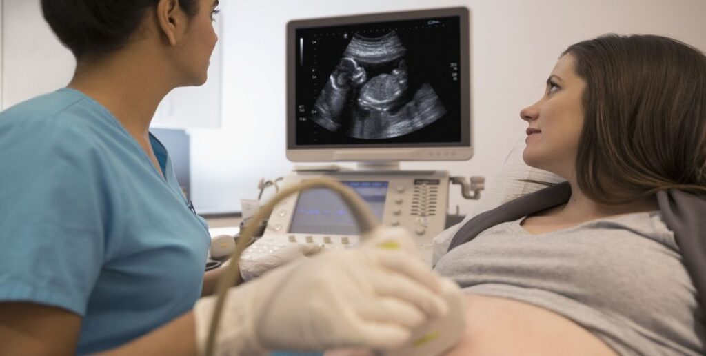
And it is also known to help monitor the pregnancy of pregnant women and ensure that the fetus is developing well and that there are no complications (miscarriage, ectopic pregnancy, etc.).
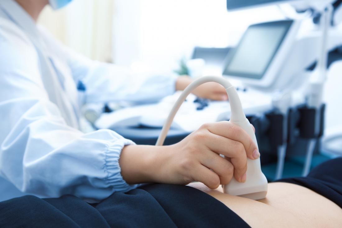
Two techniques are used. The organs can be observed through a probe placed on the skin of the belly. Ultrasound is called transcutaneous. In case of difficulties in benefiting from sufficiently clear and analyzable images, ultrasound is performed by endocavitary or endorectal route (for example for prostate ultrasound). The probe is then introduced inside the body (via the vagina or the rectum) to be as close as possible to the organs to be observed.

Cardiac echo-doppler
Cardiac echo-doppler is useful if there is a suspicion of cardiovascular disease, it makes it possible to observe several parts in motion. First of all, from the heart (cavities, valves…), but also from the vessels in charge of transporting blood to the heart and to the other organs of the body (this is the case of the aorta and the pulmonary arteries) and finally blood flow, to know how fast and in what direction the blood flows through the vessels. It is therefore used to diagnose diseases, malformations , heart failure ,arterial hypertension … It is also used to ensure follow -up after taking so-called “cardiotoxic” drugs.
It is performed by circulating a probe placed on the thorax and called “transthoracic” (ETT).
Kidney and bladder ultrasound
To explore the kidneys and the bladder, it is the kidney and bladder ultrasound that must be relied upon. It can be prescribed if the patient suffers from abdominal pain or if he is subject to urinary disorders (Presence of blood, pain). It can detect cysts, locate obstruction in the kidneys or stones for example.
Ultrasound in case of tumor
Finally, in the case of a tumor, ultrasound has the function of helping to differentiate between a cyst made up of liquid and a solid tumor, or even to know in which other areas or organs the cancer has developed. For example, thyroid cancer is asymptomatic before leading to potential lumps, difficulty breathing or swallowing. Ultrasound of the thyroid, making it possible to analyze its structure, the size of the gland, and the existence of nodules, is essential in case of doubt.
How is an ultrasound performed?
It can be practiced in many places (hospital, radiology practice, doctor’s office, etc.), as long as the room is sufficiently dark, for good visibility of the images. It does not last long, on average, 15 to 30 minutes and is ambulatory (no need to stay overnight). It can be carried out by a doctor specializing in the organ to be analyzed ( cardiologist , gynecologist, etc.), a radiologist or, in certain cases, by other health professionals.
Before the ultrasound, procedures may be required to make the organ easier to examine. In some cases, a laxative will need to be taken first. It will sometimes be necessary to come on an empty stomach, without drinking or eating, or on the contrary to drink large quantities of water.
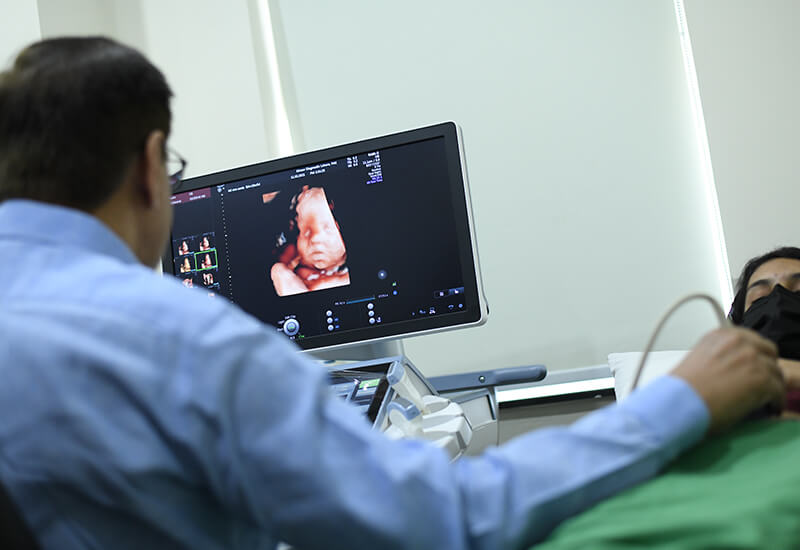
An assessment, relating to the medical history, current illnesses and medication taken, is carried out before the ultrasound. For the ultrasound itself, the patient is seated or lying down and may be asked to hold their breath. A probe, previously covered with an ultrasound-conducting gel, is positioned on the area to be analyzed (skin or by insertion into an orifice to be in direct contact with the organ). Connected to the computer, it makes it possible to transcribe moving images of the observed area using ultrasound. Some images are recorded so that the doctor can better analyze the morphology of the organs and in particular take measures. The results are generally communicated and explained after the examination, and formalized in a report, given to the patient and sometimes to the prescribing doctor.
If he deems it necessary, the doctor can recommend additional tests.

Ultrasound, is it painful?
Ultrasounds are not X-rays. The examination does not present any pain, danger or complications for the patient.
However, the pressure of the probe can be unpleasant if the area is weakened or in pain.
What documents to bring for an ultrasound?
If you are required to have an ultrasound, do not hesitate to ask your doctor’s office for the information you will need to provide on D-Day.
In particular, remember to bring your prescription, your carte Vitale, your mutual insurance card, and any other important element of your medical file (Letters from the doctor, results of examinations, reports, old x-rays, ultrasounds, etc.).
What if you get a second opinion?
Have you received ultrasound results, discussed them with your doctor, but still have questions about the diagnosis made and the procedure to follow? A second opinion from a doctor specializing in your illness may be useful to reassure you.


