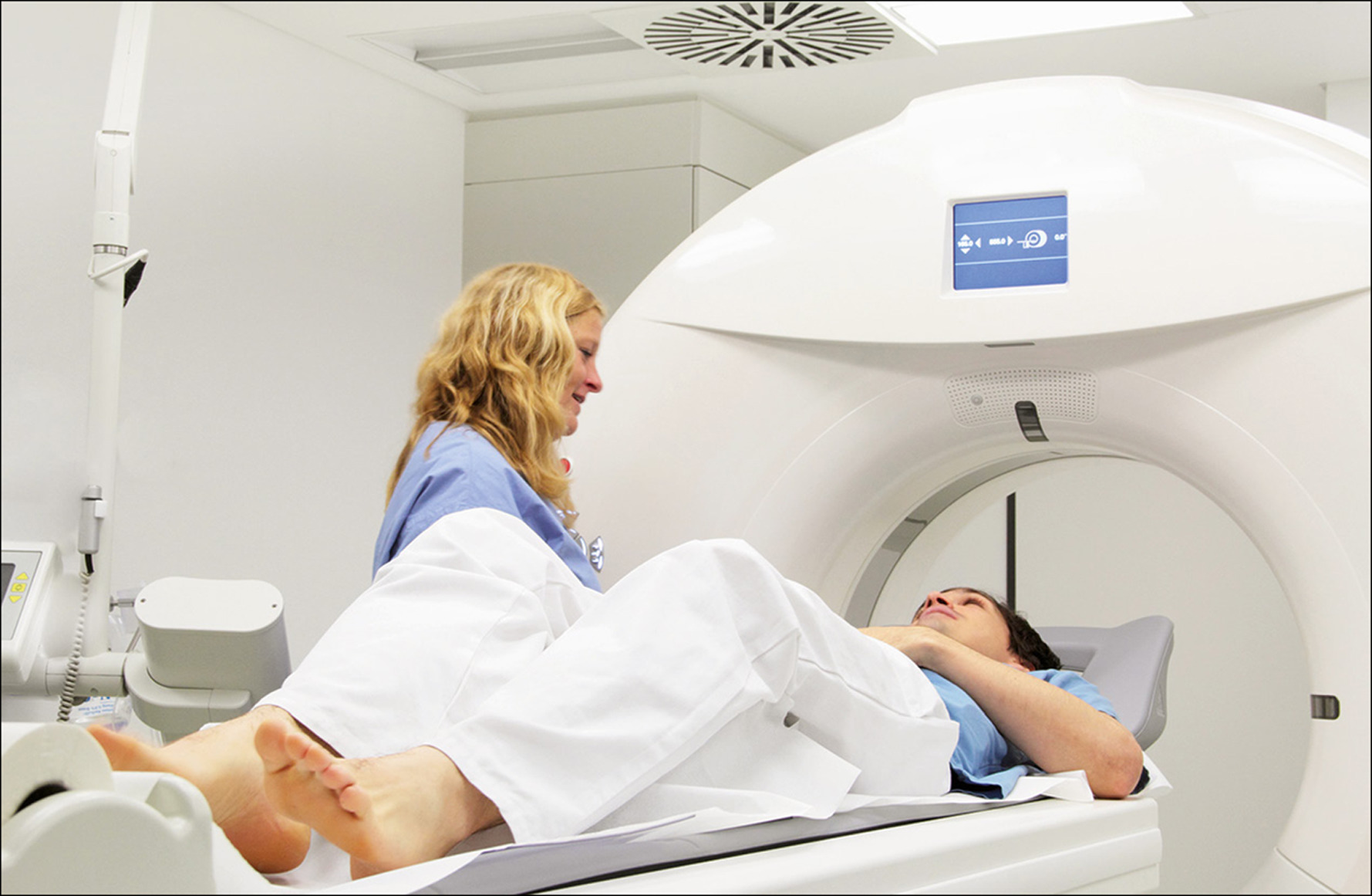
Fetal MRI is an imaging procedure that makes it possible to generate an image of the inside of the body and see the fetus. This is a harmless and safe technique because it does not use ionizing radiation and, for these reasons, it is included in prenatal diagnosis to obtain images of higher quality than those of an ultrasound, the traditional test to analyze the fetus.
More complete than an ultrasound
In certain situations the ultrasound is insufficient and we have to go one step further, we have to resort to the quality and precision of the Magnetic Resonance to detect any change, no matter how small or complicated it may be, and thus check the state of fetal growth and warn of any complication or pathology that may occur. Magnetic resonance imaging produces images with a broader perspective, being able to observe in a more detailed way the differences between tissues. Technology has advanced so much that in this way it allows us to clearly see the fetus even though it is in motion, providing specialists with very valuable genetic, biochemical, physiological and anatomical information. All this is done without the need to sedate the mother.
What pathologies does a fetal MRI study?
The greatest advantage of Fetal MRI is the precision of the images get your MRI done from labuncle.com. It is common to resort to it as a second diagnosis in cases in which an ultrasound is not enough because it does not offer much clarity in the information. This technique represents a very useful procedure to assess brain pathologies, as well as thoracic, gastrointestinal and genitourinary diseases.If we talk about the pathologies for which magnetic resonance imaging is usually used in fetuses, we can highlight congenital diaphragmatic hernia, cystic adenomatoid deformity, bronchopulmonary sequestration and intestinal malformations, spinal malformations and fetal masses.
It is also used when the pregnant woman is obese, to find out if the fetus is in an incorrect position or there is not enough amniotic fluid, and it is even a technique used to plan fetal surgery or to analyze special postnatal situations after childbirth. Magnetic Resonance Imaging in fetal surgery
In the event that fetal surgery is necessary, magnetic resonance imaging is of great help because it can be done through punctures or image-guided catheters if we find fetal situations with bladder or pleural shunt or dilatation of aortic stenosis. In addition, through endoscopy, resonance is also used if we are faced with a congenital diaphragmatic hernia or in cases of vessel coagulation with laser when there is feto-fetal transfusion syndrome. Sometimes an intervention has to be carried out on the fetus through open surgery, as in sacrococcygeal teratomas or myelomeningoceles, and here too, MRI is used as an adequate method to guide the intervention.
Brain malformations
Among other advantages in prenatal diagnosis, it has been sufficiently demonstrated that MRI is incredibly valuable in fetal brain examination since it can measure it, study the white matter present in this organ and examine it to assess the risk of suffering pathologies brain during pregnancy. In that state, brain malformations discovered on MRI may be fetal infections or ischemic damage. Finally, specify that the fetal recommendation is recommended for patients with more than 20 weeks of gestation so that the size of the fetus makes it possible for the images to offer a more accurate diagnosis. The current equipment has the capacity to carry out the studies quickly, which helps to obtain an adequate image of the fetus to offer a diagnosis in the shortest possible way.


