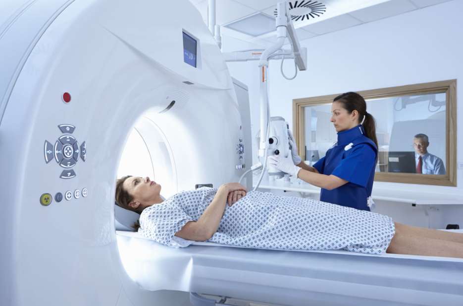
CAs in the case of MRI, CT achieves images with high temporal and spatial resolution, which makes it an ideal technique for studying the morphology of vessels, their walls, surgical anastomoses, the airway, the lung parenchyma, and sometimes assessment of ventricular function.
Indications
The main indications for performing CT in patients with congenital heart disease include:
Evaluation of the coronary vessels: it not only evaluates their outlet and proximal path, but also their distal path, calcium deposits on their walls (calcium quantification or Calcium-Score) and is a better method than MRI to assess the complications secondary to surgical procedures where they are manipulated as in the Transposition of the Great Arteries.
Evaluation of patients repaired with stents or embolization coils
Patients with pacemakers, those not compatible with MRI.
Evaluation of vascular rings, since it better evaluates their relationship with the rest of the mediastinal structures, including the airway and the esophagus.
The indication in patients with claustrophobia is relative. In my opinion, with patience and adequate preparation they would withstand the MRI. If this is not possible, consideration should be given to performing an MRI with sedation, since in any case, CT, despite current technical advances, still involves a high dose of radiation get your tests from Lab Uncle.
In patients in a very serious clinical situation that is difficult to control, or in immediate post-surgical situations who need a rapid study (5-10 minutes).
Advantages and disadvantages of CT in relation to MRI
Advantages:
A shorter scan time. In children it allows less sedation / anesthesia to be used
It is compatible in patients with pacemakers and metallic devices.
Greater availability.
Simultaneous assessment of the heart and lung with their airways.
Disadvantages
High doses of radiation. It is the main limiting factor for performing a CT, since they will need multiple evolutionary controls throughout their lives and the radiation dose is added test after test.
Need for intravenous contrast. In CT, unlike MRI, it will always be necessary to administer intravenous contrast. The contrast used in CT is based on the chemical element iodine.
Less functional information (ventricular function) than MRI
In addition, vascular calcifications or surgical material can distort the image and make it difficult to see the lumen of the vessel well. Pacemakers and other metallic devices can also produce artifacts, although in the latest generation CT, all these artifacts are minor.
Also Read: Computed Tomography (CT): what is it for, advantages and disadvantages
Examples of images obtained in the CT
In the upper images on the left, we observe the small caliber of the (hypoplastic) aortic arch (arrow) and an enlarged ductus () in a 5-day-old patient with suspected aortic arch interruption. In the upper images on the right, we observe a complete double aortic arch, with branch asymmetry, the right side being larger in caliber () in a 6-year-old patient with respiratory symptoms. The 3D reconstruction of the airway shows the stenosis it produces in the tracheal lumen (*).
In the upper images on the left, we can see a coarctation of the aorta treated with a stent in MIP projection and its VRT-3D reconstruction in a 16-year-old patient.
In the upper images on the right we see a stenosis of the pulmonary veins (arrow) in a 1-year-old child (left) and a large ASD (right)
In the upper image on the left we observe a left coronary artery aneurysm and partially calcified wall (arrow) in an 8-year-old patient with Kawasaki disease.
In the upper image on the right we observe an anomalous origin of the left coronary artery in the pulmonary trunk and subsequent correction, with reimplantation in the aorta using a Dacron tube (Arrow) in a 3-year-old girl.


